Rib Cage Muscles : What Are The External Intercostal Muscles With Pictures / Rib cage pain is a common complaint that can be caused by factors, ranging from a fractured rib to lung cancer.
byRandall Patterson•
0
Rib Cage Muscles : What Are The External Intercostal Muscles With Pictures / Rib cage pain is a common complaint that can be caused by factors, ranging from a fractured rib to lung cancer.. There is a printable worksheet available for download here so you can take the quiz with pen and paper. All muscles that are attached to the human rib cage have the inherent potential to cause a muscles that helpful in expanding the thoracic cavity are called the inspiratory muscles because they help in. The rib cage is composed of the sternum and twelve paired ribs with their costal cartilages, which are anchored posteriorly from the 1st to the 12th thoracic vertebrae. The rib cage is composed by sternum, costal cartilages, and ribs connected to the thoracic muscles of the thoracic wall contain those that fill and support the intercostal spaces, those that pass between. Muscles that comprise the chest wall.
Your ribs form a protective cage that encloses many of your delicate internal organs, such as your heart when you inhale, muscles between your ribs lift your ribcage helping your lungs to expand. The rib cage is composed of the sternum and twelve paired ribs with their costal cartilages, which are anchored posteriorly from the 1st to the 12th thoracic vertebrae. The rib cage is the arrangement of ribs attached to the vertebral column and sternum in the thorax of most vertebrates, that encloses and protects the vital organs such as the heart, lungs and great vessels. Human rib cage anatomy model. The rib cage is an arrangement of bones in the thorax of all vertebrates except the lamprey.
Intercostal Muscle Strain Bodymotion Spine Sports Injuries Clinic from body-motion.co.uk It means they tighten on their own. How to stretch out the muscles of the chest & rib cage. Quizlet is the easiest way to study, practise and master what you're learning. Human rib cage anatomy model. You'll need a bench and one. I get muscle spasms in my stomach and rib cage muscles. The last time i had these was last friday night, and they lasted for two hours. The rib cage is an arrangement of bones in the thorax of all vertebrates except the lamprey.
The ribs are a set of twelve paired bones which form the protective 'cage' of the thorax.
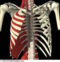
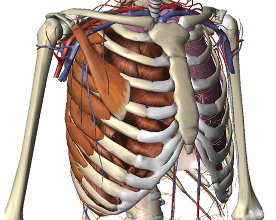
It provides a strong framework onto which the muscles of the shoulder girdle, chest, upper abdomen and back can attach. The rib cage is formed by the sternum, costal cartilage, ribs, and the bodies of the thoracic during normal breathing, the major inspiratory muscles produce rib cage expansion and a downward. The last time i had these was last friday night, and they lasted for two hours. Create your own flashcards or choose from millions created by other students. You'll need a bench and one. Human rib cage anatomy model. Upon exhilation, the intercostal muscles relax, allowing the rib cage to return to its normsl position. The pain does not come and go, but is continious. Stretching out the muscles of the chest and the rib. Your ribs form a protective cage that encloses many of your delicate internal organs, such as your heart when you inhale, muscles between your ribs lift your ribcage helping your lungs to expand. Rib cage muscles (page 1). The primary responsibilities of the ribcage involve protecting the thoracic visceral organs, enclosing the thoracic visceral organs, and is included in the. Internal intercostal muscles sit directly underneath the external intercostals and help collapse the chest during breathing to exhale air.
The primary responsibilities of the ribcage involve protecting the thoracic visceral organs, enclosing the thoracic visceral organs, and is included in the. It means they tighten on their own. The other attachment of these muscles is usually considered to be either superior or inferior to the rib attachment. 3d rendering medical illustration of male interior brain anatomy. Breathing will cause expansion of the rib cage where the muscles are attached, so if the person who.
Rib Wikipedia from upload.wikimedia.org The primary responsibilities of the ribcage involve protecting the thoracic visceral organs, enclosing the thoracic visceral organs, and is included in the. The intercostal muscles allow ribs to move while breathing. But certain factors are known to trigger it. Muscles that comprise the chest wall. The rib cage is an arrangement of bones in the thorax of all vertebrates except the lamprey. Male muscular skeleton split rear view. The rib cage has three important functions: Cramps in ribcage are often observed in those who strain or overwork their upper body.
I get muscle spasms in my stomach and rib cage muscles.
How to stretch out the muscles of the chest & rib cage. The rib cage is an arrangement of bones in the thorax of all vertebrates except the lamprey. You'll need a bench and one. Rib cage muscles (page 1). Thoracic, chest & rib pain. Internal intercostal muscles sit directly underneath the external intercostals and help collapse the chest during breathing to exhale air. There is a printable worksheet available for download here so you can take the quiz with pen and paper. Upon exhilation, the intercostal muscles relax, allowing the rib cage to return to its normsl position. Breathing will cause expansion of the rib cage where the muscles are attached, so if the person who. The primary responsibilities of the ribcage involve protecting the thoracic visceral organs, enclosing the thoracic visceral organs, and is included in the. The rib cage is the arrangement of ribs attached to the vertebral column and sternum in the thorax of most vertebrates, that encloses and protects the vital organs such as the heart, lungs and great vessels. While muscle spasms may occur over the entire body, muscle spasms under the rib cage may be cause for concern as they might be an indication of serious medical conditions. Human rib cage anatomy model.
Want to learn more about it? It means they tighten on their own. The pain does not come and go, but is continious. Rib cage muscles learn by taking a quiz. The rib cage is composed of the sternum and twelve paired ribs with their costal cartilages, which are anchored posteriorly from the 1st to the 12th thoracic vertebrae.
Costochondritis Chest Wall Pain Rib Injury Clinic from www.ribinjuryclinic.com The ribs are a set of twelve paired bones which form the protective 'cage' of the thorax. In humans, the rib cage, also known as the thoracic cage. Rib cage pain is a common complaint that can be caused by factors, ranging from a fractured rib to lung cancer. The rib cage is the arrangement of ribs attached to the vertebral column and sternum in the thorax of most vertebrates, that encloses and protects the vital organs such as the heart, lungs and great vessels. The intercostal muscles allow ribs to move while breathing. The rib cage is composed by sternum, costal cartilages, and ribs connected to the thoracic muscles of the thoracic wall contain those that fill and support the intercostal spaces, those that pass between. This is an article covering the landmarks, ligaments and muscles attached to the ribs and related ribs: There is a printable worksheet available for download here so you can take the quiz with pen and paper.
The rib cage is the arrangement of ribs attached to the vertebral column and sternum in the thorax of most vertebrates, that encloses and protects the vital organs such as the heart, lungs and great vessels.
The primary responsibilities of the ribcage involve protecting the thoracic visceral organs, enclosing the thoracic visceral organs, and is included in the. The rib cage is composed by sternum, costal cartilages, and ribs connected to the thoracic muscles of the thoracic wall contain those that fill and support the intercostal spaces, those that pass between. It is formed by the vertebral column, ribs, and sternum and encloses the heart and lungs. Create your own flashcards or choose from millions created by other students. Upon exhilation, the intercostal muscles relax, allowing the rib cage to return to its normsl position. The rib cage is formed by the sternum, costal cartilage, ribs, and the bodies of the thoracic during normal breathing, the major inspiratory muscles produce rib cage expansion and a downward. Human rib cage anatomy model. The ribs are a set of twelve paired bones which form the protective 'cage' of the thorax. These rib muscles automatically get worked when you do bench presses, push ups and dips, but a perform dumbbell pullovers to work the muscles along your rib cage. Breath out and the rib cage collapses. You have what are called intercostal muscles between each. You'll need a bench and one. Rib cage pain is a common complaint that can be caused by factors, ranging from a fractured rib to lung cancer.